
A fluorescence microscope is a powerful equipment used to observe and study biological material at the cellular and molecular levels in scientific research, medical diagnostics, and a variety of other fields. Opposed to ordinary microscopes that rely on transmitted or reflected light, it uses the special features of fluorescent molecules to produce colorful, high-contrast images. In this article, we’ll take you through the topic of how does a fluorescence microscope work, exploring the inner workings of a fluorescence microscope and understanding how it harnesses the phenomenon of fluorescence to reveal intricate details in biological samples.
What is the Basic Working Principle of a Fluorescence Microscope?
A fluorescence microscope’s primary principle is based on the phenomenon of fluorescence, which is the emission of light by certain molecules when activated by a specific wavelength of light. Here’s a summary of fluorescence microscopy’s fundamental principles:
- Fluorescent Molecules
Fluorescence microscopy makes use of fluorescent molecules, also known as fluorophores, which are dyes or chemicals that can absorb light of a given wavelength and then emit light of a longer wavelength. These fluorophores can be naturally occurring or synthetic, and they come in a variety of colors, each emitting light at distinct wavelengths.
- Excitation and Emission
Fluorescence involves two major steps: excitation and emission. When a sample containing fluorescent molecules is exposed to a specific wavelength of excitation light, the fluorophores absorb this energy and are promoted to a higher energy state. This higher state, however, is unstable, and the fluorophores quickly return to their original, lower energy state, releasing the surplus energy as light.
- Specific Wavelengths
Each fluorophore has its own excitation and emission wavelengths, which are critical for fluorescence microscopy. The excitation wavelength is the amount of light needed to excite the fluorophore, while the emission wavelength is the amount of light released as a result of this excitation.
- Selective Detection
A fluorescence microscope is outfitted with filters that allow for the selective detection of fluorescent light emitted. Only the emitted fluorescence, which has a longer wavelength, is permitted to pass through to the detector once the excitation light is filtered off. This selective detection ensures that the image captured is unique to the fluorophores in the sample.
- High Contrast and Sensitivity
Because of its capacity to detect emitted light selectively, fluorescence microscopy provides excellent contrast and sensitivity. This allows researchers to differentiate fluorescently tagged structures from the background and see small details within the specimen.
- Labeling Biological Samples
Biological samples are frequently labeled with fluorescent markers or dyes that target specific cellular structures, proteins, or molecules of interest in fluorescence microscopy. Multiple structures can be tagged and observed simultaneously in the same sample by utilizing separate fluorophores with varied emission colors, allowing for multi-color imaging.
- Applications
Fluorescence microscopy is utilized extensively in a variety of scientific disciplines, including cell biology, immunology, neurology, microbiology, genetics, and medicine. It enables researchers to use high accuracy and real-time imaging to examine cellular processes, protein localization, interactions, and dynamics.
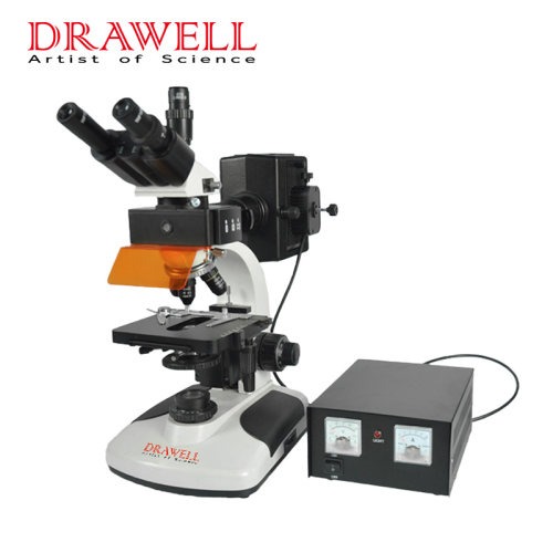
What are the Key Components of a Fluorescence Microscope?
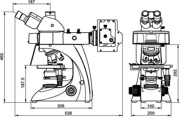
These key components work together to excite and detect fluorescence in a fluorescence microscope.
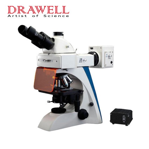
- Light Source
A fluorescence microscope’s light source is typically a high-intensity lamp or a laser. To excite the fluorophores in the sample, it generates strong light of a specified wavelength. The light source utilized is determined by the excitation wavelength required for the specific fluorophores used.
- Excitation Filter
The excitation filter is used to separate the light source from the sample. It selectively transmits the chosen wavelength of excitation light while blocking other wavelengths from reaching the sample. Only the excitation light capable of activating the fluorophores is directed onto the specimen by this filter.
- Dichroic Mirror
A dichroic mirror, commonly known as a beam splitter, is an important part of fluorescence microscopy. It reflects the excitation light towards the sample while allowing the fluorescence emitted to pass through. The dichroic mirror is intended to have a high reflectivity at the excitation wavelength and a high transmittance at the longer emission wavelength.
- Objective Lens
The objective lens is in charge of gathering and focusing the excitation light on the sample. It also collects and focuses the light generated by the fluorophores back onto the detector. The quality of the objective lens has a considerable impact on picture resolution and overall microscope performance.
- Emission Filter
Between the sample and the detector is the emission filter, also known as the barrier filter. It permits only the required wavelength of emitted fluorescence to pass through while preventing any residual excitation light and scattered light from reaching the detector. Only the specified fluorescence signal is recognized by this filter.
- Detector
The detector collects the fluorescence generated by the emission filter. Photomultiplier tubes (PMTs) and charged-coupled devices (CCDs) are common detectors. PMTs are extremely sensitive and can detect even the faintest fluorescent signals, making them excellent for low-light applications. CCDs, on the other hand, provide high-resolution imaging and are frequently employed for digital image capture.
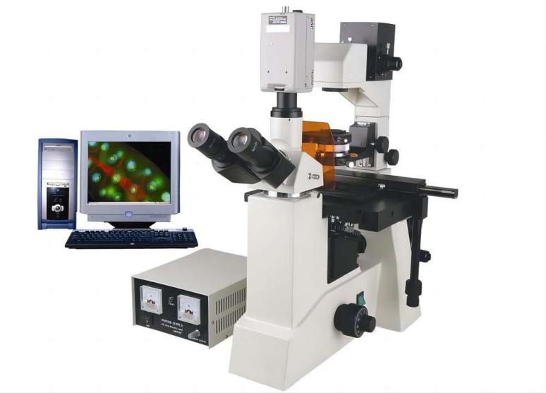
- Camera or Imaging System
The detector output is frequently coupled to a camera or image system in modern fluorescence microscopes. This enables the observation and digital recording of fluorescence images in real-time. High-quality cameras can provide detailed, high-resolution photos that can be used for a variety of applications.
- Fluorescent Probes
Fluorescent probes or fluorophores are crucial to fluorescence microscopy, despite the fact that they are not physical components of the microscope. When exposed to a suitable wavelength of light, these dyes or compounds emit fluorescence. Fluorophores are chemicals that are used to mark and observe specific cellular structures or molecules in a sample.
Summary
Fluorescence microscopy has transformed how scientists and researchers examine and study biological samples. Fluorescence microscopes enable visibility at the molecular level by capitalizing on the phenomenon of fluorescence, providing essential insights into the complicated world of cells and molecules.