

In materials analysis, X-ray based approaches are kinda invaluable for trying to get a grip on composition, structure, and properties, overall. Two of the most used options are X-ray Diffraction (XRD) and X-ray Fluorescence (XRF). Even though both of XRD and XRF depend on X-rays, the basic idea behind them, their actual use cases, and the kind of outputs they produce are fundamentally not the same. Figuring out these contrasts matters a lot, because choosing the right technique for a particular analytical situation becomes much easier when you know what each one really tells you.
Understanding XRD
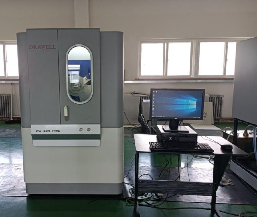
X-ray Diffraction( XRD) is a potent analytical approach that shows up everywhere in materials science, chemistry, geology, and engineering, to peek at the atomic and molecular makeup of crystalline substances. When X-rays hit a crystal, they get scattered, and the overall behavior of that scattering helps reveal what the atoms are doing. Because of that, XRD gives useful information that matters both for research efforts and for industrial use.
Principles
XRD leans on X-ray diffraction itself, meaning, when X-rays hit a crystalline material, they scatter into particular directions. That scattering is not random, it forms a diffraction pattern, and this pattern is tied to how atoms are arranged across the crystal lattice. Then by looking at the diffraction angles and the relative strengths, researchers can infer the crystal structure, distinguish phases, estimate lattice parameters, and even see signs of structural defects or lattice strain inside the specimen.

Advantages
- Phase Identification: With XRD you can pin down crystalline phases in a material, even when the mixture is messy or complex. This is useful in mineralogy, metallurgy, and also pharmaceuticals, yes.
- Structural Analysis: XRD gives insight on crystal structure, unit cell dimensions, and how atoms are actually arranged inside. That matters a lot if you want to understand what a material can do and why.
- Non-Destructive: In many cases, the sample stays intact after the measurements, so the same specimen can be checked again later or put back to further use.
- Quantitative Phase Analysis: XRD also can estimate how much of each crystalline phase is present in a combined sample, not just name them.
- Detection of Strain and Defects: XRD can show residual stress, dislocations, and other lattice imperfections. These are vital for materials engineering
- Versatility: The method works for powders, thin films, and even bulk materials, as long as sample preparation is handled the right way.
Limitations
- Requires Crystallinity: XRD does not do well with amorphous, or non-crystalline substances, because they give broad diffraction patterns with hardly any distinct features
- Sample Preparation: Powders should be finely ground and pretty homogeneous, because surface irregularities can skew or distort the results in a not so obvious way.
- Limited Sensitivity to Light Elements: Elements like hydrogen, lithium, or beryllium are difficult to detect due to weak scattering of X-rays.
- Complex Data Interpretation: Diffraction patterns from multi-phase or highly complex materials require expert analysis and sometimes sophisticated software.
- Time-Consuming for High-Resolution Studies: Detailed structural analysis can take longer, especially when measuring small lattice distortions or textures.
- Equipment Cost: XRD instruments are expensive and require maintenance and calibration, limiting accessibility in some laboratories.
Understanding XRF
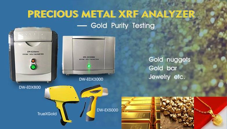
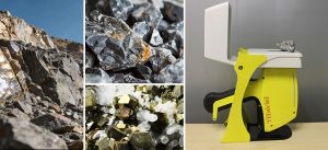
X-ray Fluorescence(XRF) is a widely used analytical method for figuring out the elemental make up of materials. People like it because it works fast, feels straightforward, and it does not destroy the sample, so it is often picked in things like mining, metallurgy , environmental tracking , and manufacturing inspections. When you shine a primary X-ray source on a material, the sample answers by sending back characteristic secondary X-rays. With those signals XRF can give both identifying insights and measured amounts of the elements that are in the sample , at the same time.
Principles
XRF mainly relies on how energetic X-rays interact with atoms inside the sample. Once the material is hit by the primary X-rays, electrons from inner shells can be knocked out, leaving behind vacancies. Then electrons from higher energy states fall down to these missing spots, and the released energy comes out as fluorescent X-rays.
Each element, emits X-rays at particular, characteristic energy levels. This lets the instrument basically pin down which elements are actually in the material, not guessing too much. The intensity of the X-rays that come out is tied to the concentration of that element, so quantitative analysis becomes possible. Depending on how the device is built, XRF can be done with wavelength dispersive systems (WDXRF) or energy dispersive systems (EDXRF), and each one tends to bring different tradeoffs in precision and speed.
Advantages
- Non-destructive Analysis: XRF does not change or ruin the sample, so it fits well for precious or limited items.
- Rapid Results: Most measurements finish in just seconds to minutes, which helps high-throughput screening and faster choices on the spot.
- Minimal Sample Preparation: Solid pieces, powders, liquids, or pressed pellets can often be measured directly, with only slight setup.
- Wide Elemental Range: XRF can cover a broad spectrum of elements. Typically, it runs from sodium (Na) to uranium (U), depending on the instrument you use.
- Good Accuracy for Major and Minor Elements: It is highly effective for working out alloy composition, geological sample material, and industrial substance characteristics.
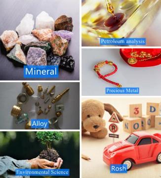
- Versatility across industries: This method gets used broadly in mining exploration, cement manufacturing, metal production, environmental checks, and regulatory compliance tasks (such as RoHS screening).
Limitations
- Limited Sensitivity when It Comes to Light Elements: Hydrogen, lithium, and sometimes boron or carbon are difficult to measure precisely, or they may be effectively impossible to detect.
- No Structural Information: XRF points to elemental makeup only ; it cannot tell you anything about crystal structure, bonding types , or the specific phases present.
- Matrix Effects: Sample composition can influence X-ray absorption and the emission pattern, so calibration plus corrections are needed for dependable numbers.
- Surface Sensitivity: XRF mostly looks at near-surface layers, so for heterogeneous samples it might miss what the bulk composition looks like.
- Detection Limits for Trace Elements: Even when the approach works well for major and less concentrated elements, the extremely tiny trace amounts can be hard to capture if you do not have advanced instrumentation available.
- Calibration Dependency: Accurate quantitative results depend on having standards that are prepared properly, and on reliable calibration models, especially when the sample matrix is complex.
Key Differences Between XRD and XRF
| Aspect | XRD | XRF |
| Primary Information | Crystal structure, phase identification | Elemental composition |
| Material Type | Crystalline materials | Both crystalline and amorphous |
| Sample Preparation | Requires fine powder and uniform surface | Minimal, can analyze solids, powders, liquids |
| Analysis Speed | Moderate (minutes to hours) | Fast (seconds to minutes) |
| Non-destructive | Generally non-destructive | Non-destructive |
| Detection of Light Elements | Limited | Very limited |
| Structural Information | Provides detailed atomic arrangement | Not provided |
| Quantitative Analysis | Phase quantification possible | Elemental quantification possible |
| Applications | Mineralogy, metallurgy, pharmaceuticals | Mining, quality control, environmental testing |

Key Factors to Consider for Choosing Between XRD and XRF
Selecting between XRD and XRF depends on a careful evaluation of the analytical needs.
1. Understanding the Analytical Goal
First step when you pick between XRD and XRF is to lock in the analytical objective first. XRD is mainly used for phase identification and crystallographic analysis, giving more granular data about the crystal structure, lattice parameters, and the mineral phases inside your sample. That is why it works well for research, quality control of crystalline materials, and studies that truly need structural insights.
XRF, however, is built for elemental composition analysis, able to identify and quantify a large range of elements, from sodium up to uranium. XRF shows up a lot in geology, environmental monitoring, metallurgy, and industrial use cases where the main focus is on determining elemental concentrations.
2. Sample Type and Preparation
Sample characteristics can heavily influence the choice. XRD requires a well-prepared, homogeneous sample with sufficient crystallinity for accurate diffraction measurements. Powdering, grinding, or pressing the sample into a pellet is often necessary, which may be time-consuming for some materials.
XRF is generally more flexible, accommodating powders, liquids, solids, and even coatings with minimal preparation. This makes it advantageous when rapid analysis or non-destructive testing is required.
3. Detection Limits and Sensitivity
Another critical factor is the detection sensitivity for the elements or phases of interest. XRF offers high sensitivity for medium to heavy elements, but its detection of light elements like lithium, beryllium, or boron can be limited. XRD does not provide direct elemental concentrations but can detect minor or trace crystalline phases if they are present in sufficient amounts.
For uses where trace element quantification matters a lot, XRF is often the more preferred way, yes. When the emphasis shifts to picking out minor phases, or crystalline patterns in a substance, then XRD tends to fit better, in practice.
4. Quantitative vs Qualitative Analysis
XRF does very well at quantified elemental work, giving accurate concentration values, using calibration that is usually pretty straightforward. XRD can do semi-quantified phase analysis, for example via Rietveld refinement, but it is commonly less reliable if you need direct elemental quantity.
So if the purpose is to measure exact elemental concentrations, XRF gives the advantage. If the purpose is to learn about crystal structure, phase makeup, or lattice details then XRD becomes the method of choice.
5. Analysis Speed and Throughput
Selection can be influenced by operational efficiency as well. XRF usually gives faster results and a high throughput, which makes it fitting for industrial quality control, or environmental monitoring in practice. XRD experiments often take more time for the setup and the measurement part, especially if you need detailed structural characterization, or phase refinement rather than a simple scan.
6. Cost and Accessibility
The cost of the equipment , and the overall operational complexity also matter. In general, XRD instruments are more expensive, they call for skilled operators , and they tend to have longer maintenance cycles. XRF setups can run from benchtop portable devices up to high-end laboratory instruments, so there is usually more flexibility regarding budget and access.
7. Environmental and Operational Considerations
There are applications where you really need non-destructive testing or the ability to measure samples in situ, without messing with them too much. In these situations XRF is usually the choice, because it allows minimal sample preparation and stays non-destructive. Now XRD is also non-destructive in principle, but in practice it often asks for additional preparation steps, and that preparation can end up changing, consuming, or otherwise affecting the sample content.
Summary
- Selecting XRD when structural characterization, phase identification, or crystallographic analysis is essential.
- Choosing XRF when elemental composition, rapid analysis, minimal sample preparation, and quantitative measurements are the priority.

Final Thoughts
Selecting between XRD and XRF depends on the type of information needed. In many industrial and research contexts, both techniques are complementary, providing a comprehensive understanding of both structure and composition. By recognizing the strengths and limitations of XRD and XRF, scientists and engineers can make informed decisions that optimize analysis, improve material development, and ensure quality control.
Get Quote Here!
Latest Posts


What Next?
For more information, or to arrange an equipment demonstration, please visit our dedicated Product Homepage or contact one of our Product Managers.
For more information, or to arrange an equipment demonstration, please visit our dedicated Product Homepage or contact one of our Product Managers.


