
Fluorescence microscopy is a type of optical microscope that uses fluorescence and phosphorescence instead of or in addition to reflection and absorption to study the properties of organic or inorganic substances. Fluorescence is the emission of light from substances that absorb light or other electromagnetic radiation, while phosphorescence is a specific type of photoluminescence related to fluorescence. Unlike fluorescence, a phosphorescent material does not immediately re-emit the radiation it absorbs. Fluorescence microscopy was invented in the early twentieth century by August Kohler, Karl Reichert, and Heinrich Lehmann, among others.
Principles of Fluorescence Microscopy
Most cellular components are colorless and indistinguishable under a microscope. The basic premise of fluorescence microscopy is to stain parts with dyes.
Fluorescent dyes, also known as fluorophores or fluorochromes, are molecules that absorb excitation light at a given wavelength (usually UV). After a short delay, longer wavelength light is emitted. The delay between absorption and emission is negligible, typically on the order of nanoseconds.
Emission light can then be filtered from excitation light to reveal the location of the fluorophore.
Fluorescence microscopes use much higher intensity light to illuminate the sample. This light excites fluorescent substances in the sample, which then emit light at longer wavelengths.
The resulting image is based on the emission wavelength of the secondary light source or fluorophore, rather than from the light originally used to illuminate and excite the sample.
Working with Fluorescence Microscopy
Light at the excitation wavelength is focused on the specimen through the objective lens. The fluorescence emitted by the specimen is focused on the detector through the objective lens. Since most of the excitation light is transmitted through the sample, only the reflected excitation light reaches the objective along with the emitted light.
Fluorescence microscopy imaging modality
“Fluorescence microscope” refers to any microscope that uses fluorescence to generate images, whether it is a simpler setup like an epifluorescence microscope, or a more complex design like a confocal microscope, which uses optical sectioning for better resolution Fluorescence image.
Most fluorescence microscopes used are epifluorescence microscopes, in which the excitation of fluorophores and detection of fluorescence is accomplished through the same optical path (i.e. through the objective lens).
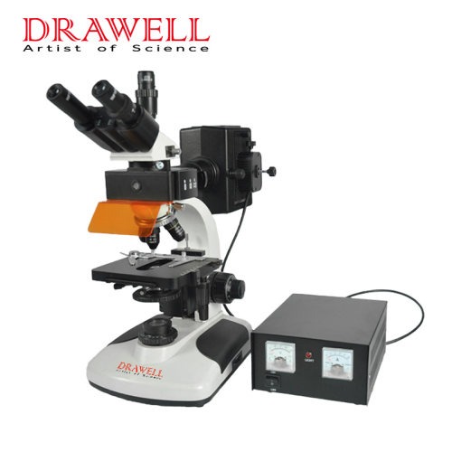
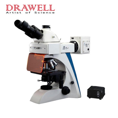
The components of a fluorescence microscope
a fluorescence microscope consists of Fluorescent dyes (fluorophores), a light source, dichroic mirror, Launch filter
Fluorescent dyes (fluorophores)
A fluorophore is a fluorescent compound that re-emits light when excited by light.
Fluorophores usually contain several combined aromatic groups or are planar or cyclic molecules with multiple π-bonds.
Many fluorescent dyes are designed for a range of biomolecules.
Some of these are small molecules that are inherently fluorescent and bind biomolecules of interest. Prime examples of these are nucleic acid stains such as DAPI and Hoechst, and phalloidin for staining actin fibers in mammalian cells.
A light source
Four main types of light sources are used, including xenon arc lamps or mercury vapor lamps with excitation filters, lasers, and high-power LEDs.
Lasers are mainly used in complex fluorescence microscopy techniques, while xenon lamps, mercury lamps, and LEDs with dichroic excitation filters are often used in widefield epi-fluorescence microscopy.
Excitation filter
Exciters are typically bandpass filters that pass only the wavelengths absorbed by the fluorophore, thereby minimizing excitation from other sources of fluorescence and blocking excitation light in the fluorescence emission band.
Dichroic mirror
A dichroic filter, or thin film filter, is a very precise color filter used to selectively pass light from a small range of colors while reflecting other colors.
Launch filter
The emitter is usually a bandpass filter that only passes the wavelength emitted by the fluorophore and blocks all unwanted light outside that band—especially excitation light.
Filters ensure the darkest possible background by blocking unwanted excitation energy (including UV and IR) or sample and system autofluorescence.
Applications of Fluorescence Microscopy
Identify structures in fixed and living biological samples.
Fluorescence microscopy is a common tool in life science research today as it allows the use of multicolor staining, labeling of structures within cells, and measurement of the physiological state of cells.
Advantages of Fluorescence Microscope
Fluorescence microscopy is the most popular method for studying the dynamic behavior exhibited in live-cell imaging.
This stems from its ability to isolate individual proteins with a high degree of specificity amidst non-fluorescing material.
The sensitivity is high enough to detect as few as 50 molecules per cubic micrometer.
Different molecules can now be stained with different colors, allowing multiple types of molecules to be tracked simultaneously.
These factors combine to give fluorescence microscopy a clear advantage over other optical imaging techniques, for both in vitro and in vivo imaging.