
In the realm of scientific research and diagnostics, the fluorescence microscope has revolutionized our understanding of cellular structures and processes. Among the various types of microscopes, the trinocular fluorescent microscope stands out for its versatility and advanced imaging capabilities. However, to harness the full potential of this remarkable tool, researchers and users must be aware of several crucial matters needing attention. In this article, we will delve into the best practices and precautions associated with using a trinocular fluorescent microscope, focusing on sample preparation, adjustments, maintenance, safety considerations, and image analysis.
Proper Handling and Maintenance for Using a Trinocular Fluorescent Microscope
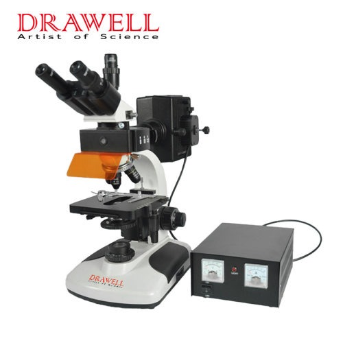
Before diving into the technical aspects, it is essential to emphasize the significance of proper handling and maintenance for the longevity and performance of your trinocular fluorescent microscope. Familiarize yourself with the microscope’s components and functions by referring to the user manual. Handle the microscope with care, avoiding rough handling or dropping it, and use lens paper or a soft cloth to clean the lenses and optical system. Remember to keep the microscope covered when not in use to prevent dust accumulation. Regular maintenance, including cleaning and replacing fluorescent bulbs, and ensuring electrical safety and proper grounding, should be conducted.
Sample Preparation Before Using a Trinocular Fluorescent Microscope
The quality of your microscopic images heavily relies on the preparation of your samples. When working with a trinocular fluorescent microscope, it is crucial to employ suitable mounting techniques to ensure proper fixation of the specimen without applying excessive pressure or squeezing. Additionally, carefully choose appropriate fluorescent dyes or markers and follow established protocols for staining. Clear labeling of the samples will aid in easy identification and accurate analysis.
Adjustments and Optimization When Using a Trinocular Fluorescent Microscope
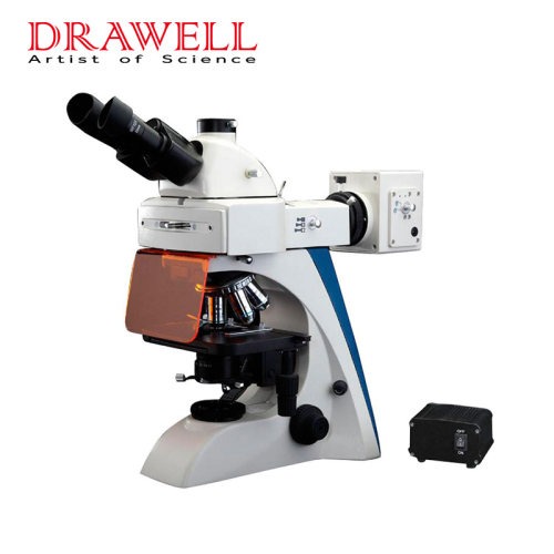
Proper alignment and optimization of the microscope are key factors in obtaining high-quality images. Begin by adjusting the interpupillary distance and aligning the binocular eyepieces to match your eyes’ comfort. Additionally, adjust the diopter settings to optimize the clarity of the observed specimen. To enhance fluorescence imaging, adjust the excitation and emission filters according to the specific dyes used. Control the intensity of the light source and optimize exposure time and gain settings for ideal image brightness and contrast.
Best Practices for Image Capture and Analysis by Using aTrinocular Fluorescent Microscope
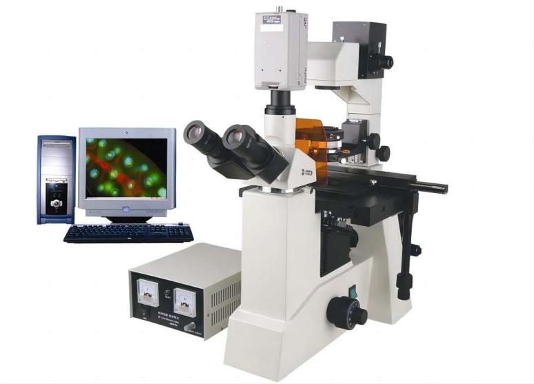
To capture and analyze your fluorescent images effectively, pay attention to the setup and configuration of the camera attached to your trinocular microscope. Ensure proper attachment and alignment, and adjust camera settings to optimize image quality. During image acquisition, capture multiple images for comparison and document experimental conditions and settings accurately. It is crucial to store and organize images and associated data securely for future reference and analysis.
Safety Considerations for Using A Trinocular Fluorescent Microscope
While exploring the wonders of fluorescence microscopy, it is imperative to prioritize safety. Always adhere to proper personal protective equipment (PPE) guidelines, ensuring the use of gloves, lab coats, and safety goggles. Handle hazardous chemicals and specimens with caution, following established protocols. Dispose of hazardous waste in compliance with relevant regulations and maintain adequate ventilation in the laboratory environment.

Conclusion
A trinocular fluorescent microscope is a powerful tool in the hands of a skilled researcher or technician. By paying attention to the matters needing attention outlined in this article, you can unlock its full potential and achieve optimal results in your experiments and analyses. Proper handling and maintenance, meticulous sample preparation, precise adjustments, adherence to safety considerations, and effective image capture and analysis are all crucial aspects to master. With dedication and attention to detail, you can navigate the fascinating world of fluorescence microscopy with confidence and achieve groundbreaking discoveries in your scientific pursuits.