

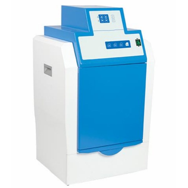
Gel imaging refers to the detection and analysis of DNA/RNA/protein and other gel electrophoresis with different stainings, such as EB, coomassie brilliant blue, silver staining, SYBRGreen, microcellular plate, plate and other non-chemiluminescence imaging. The gel documentation system can be used in conventional bioengineering research such as molecular weight calculation, density scanning, density quantification, PCR quantification, etc.
Classification of Gel Documentation System
1. The common gel documentation system
It can collect images of protein electrophoresis gel and DNA gel for qualitative analysis, including nucleic acid monitoring stained with EB, SYBRGreen, SYBRGold, TexasRed, GelStar, Fluoroscecin, RadiantRed, etc. Imaging of CoomassieBlue, SYPROOrange, various dyed protein gel, such as Kaoran, or UV, EB, and colored visible sample.
2. Chemiluminescence gel documentation system
It covers imaging of UV, EB, chemiluminescence, UV fluorescence, and colored visible sample.
3. Multicolor fluorescence gel documentation system
It covers imaging of UV, EB, chemiluminescence, multicolor fluorescence, colored visible samples, multi-functional living imaging system UV, EB, chemiluminescence, multicolor fluorescent fluorescence, tissue in vitro and animals.
Maintenance of Gel Documentation System
- The maintenance cycle of the gel documentation machine is generally about 2 weeks. The instrument shall be kept clean. Open the instrument and observe whether the software data line is in normal contact and the software operation function is intact.
- The machine computer album should be dedicated to avoiding being infected with computer viruses, which will make it unusable. At the same time, it is forbidden to reinstall the system, because the instrument software needs to be reactivated before it can be used.
- The correct closing method shall be used for closing the door of the dark box. The specific process is to cover the door of the dark box to about 60 degrees and let it go. Relying on gravity, the door will close by itself. It should be noted that you should not push hard to close the door of dark box.
- During normal use and maintenance, pay attention to keeping the UV light box and projection white light plate clean, so as not to affect the gel imaging effect of the gel documentation system.
- In the process of operating the gel documentation, in addition to the correct use of the instrument, it is also necessary to strictly comply with the provisions of the laboratory. The gel documentation system can qualitatively analyze the separation and purification results of proteins, nucleic acids, polypeptides, amino acids, polyaminoacids and other biological molecules.
Precautions For Using Of Gel Documentation System
- First turn on the system, then turn on the computer and enter the software.
- When taking ultraviolet gel photography, prevent EB from polluting the instrument, and do not use contaminated gloves to contact the door of the gel documentation system.
- The front panel of the gel documentation system cannot be opened when taking photos with the ultraviolet light source.
- After taking the photo, take out the waste glue and wipe it with soft paper.
- Focus gently.
- Please use a regulated power supply.
- Keep the indoor environment dry and wipe the water or other liquid left on the observation station in time.
- When using the instrument, close the door and observation station tightly, otherwise the ultraviolet lamp can’t be used normally.
- Don’t connect the computer to the Internet or LAN as much as possible, and install antivirus software on the computer simultaneously.
- When the instrument is not used for a long time, please cover it with a dust cover.
- To extend the service life of the lamp, please turn off the light source timely after observing the gel.


